Elbow X-Ray Normal . On the lateral image there is often a visible triangle of low density lying anterior to the. Soft tissue areas, cortical margins, trabecular patterns, bony alignment, joint. The ap view should be obtained with the elbow fully extended, the forearm fully supinated (if possible), and the fingers slightly flexed. — standard radiographic examination of the elbow includes anteroposterior (ap) and lateral views. — the elbow series is a set of radiographs taken to investigate elbow joint pathology, often in the context of trauma. Your technologist will return to reposition your elbow as needed. Normal bones and joint alignment. a recommended systematic checklist for reviewing musculoskeletal exams is:
from mavink.com
— the elbow series is a set of radiographs taken to investigate elbow joint pathology, often in the context of trauma. On the lateral image there is often a visible triangle of low density lying anterior to the. — standard radiographic examination of the elbow includes anteroposterior (ap) and lateral views. Your technologist will return to reposition your elbow as needed. Soft tissue areas, cortical margins, trabecular patterns, bony alignment, joint. Normal bones and joint alignment. a recommended systematic checklist for reviewing musculoskeletal exams is: The ap view should be obtained with the elbow fully extended, the forearm fully supinated (if possible), and the fingers slightly flexed.
Right Elbow X Ray Anatomy
Elbow X-Ray Normal Soft tissue areas, cortical margins, trabecular patterns, bony alignment, joint. a recommended systematic checklist for reviewing musculoskeletal exams is: On the lateral image there is often a visible triangle of low density lying anterior to the. Normal bones and joint alignment. Your technologist will return to reposition your elbow as needed. — the elbow series is a set of radiographs taken to investigate elbow joint pathology, often in the context of trauma. The ap view should be obtained with the elbow fully extended, the forearm fully supinated (if possible), and the fingers slightly flexed. — standard radiographic examination of the elbow includes anteroposterior (ap) and lateral views. Soft tissue areas, cortical margins, trabecular patterns, bony alignment, joint.
From
Elbow X-Ray Normal Normal bones and joint alignment. Soft tissue areas, cortical margins, trabecular patterns, bony alignment, joint. — the elbow series is a set of radiographs taken to investigate elbow joint pathology, often in the context of trauma. — standard radiographic examination of the elbow includes anteroposterior (ap) and lateral views. a recommended systematic checklist for reviewing musculoskeletal exams. Elbow X-Ray Normal.
From
Elbow X-Ray Normal — the elbow series is a set of radiographs taken to investigate elbow joint pathology, often in the context of trauma. The ap view should be obtained with the elbow fully extended, the forearm fully supinated (if possible), and the fingers slightly flexed. On the lateral image there is often a visible triangle of low density lying anterior to. Elbow X-Ray Normal.
From
Elbow X-Ray Normal a recommended systematic checklist for reviewing musculoskeletal exams is: Soft tissue areas, cortical margins, trabecular patterns, bony alignment, joint. — standard radiographic examination of the elbow includes anteroposterior (ap) and lateral views. On the lateral image there is often a visible triangle of low density lying anterior to the. Your technologist will return to reposition your elbow as. Elbow X-Ray Normal.
From www.slideshare.net
Pediatric Elbow Examination and Xrays Elbow X-Ray Normal Normal bones and joint alignment. Soft tissue areas, cortical margins, trabecular patterns, bony alignment, joint. — the elbow series is a set of radiographs taken to investigate elbow joint pathology, often in the context of trauma. On the lateral image there is often a visible triangle of low density lying anterior to the. a recommended systematic checklist for. Elbow X-Ray Normal.
From www.pinterest.co.kr
Elbow radiography. Elbow Xray view from radiology imagery , Ad, ray Elbow X-Ray Normal Soft tissue areas, cortical margins, trabecular patterns, bony alignment, joint. a recommended systematic checklist for reviewing musculoskeletal exams is: The ap view should be obtained with the elbow fully extended, the forearm fully supinated (if possible), and the fingers slightly flexed. — standard radiographic examination of the elbow includes anteroposterior (ap) and lateral views. Normal bones and joint. Elbow X-Ray Normal.
From
Elbow X-Ray Normal The ap view should be obtained with the elbow fully extended, the forearm fully supinated (if possible), and the fingers slightly flexed. Soft tissue areas, cortical margins, trabecular patterns, bony alignment, joint. — the elbow series is a set of radiographs taken to investigate elbow joint pathology, often in the context of trauma. On the lateral image there is. Elbow X-Ray Normal.
From
Elbow X-Ray Normal Your technologist will return to reposition your elbow as needed. Soft tissue areas, cortical margins, trabecular patterns, bony alignment, joint. a recommended systematic checklist for reviewing musculoskeletal exams is: — the elbow series is a set of radiographs taken to investigate elbow joint pathology, often in the context of trauma. Normal bones and joint alignment. The ap view. Elbow X-Ray Normal.
From www.aliem.com
normalelbowlateral ALiEM Elbow X-Ray Normal Your technologist will return to reposition your elbow as needed. — standard radiographic examination of the elbow includes anteroposterior (ap) and lateral views. a recommended systematic checklist for reviewing musculoskeletal exams is: — the elbow series is a set of radiographs taken to investigate elbow joint pathology, often in the context of trauma. On the lateral image. Elbow X-Ray Normal.
From
Elbow X-Ray Normal — the elbow series is a set of radiographs taken to investigate elbow joint pathology, often in the context of trauma. On the lateral image there is often a visible triangle of low density lying anterior to the. a recommended systematic checklist for reviewing musculoskeletal exams is: Soft tissue areas, cortical margins, trabecular patterns, bony alignment, joint. Normal. Elbow X-Ray Normal.
From
Elbow X-Ray Normal The ap view should be obtained with the elbow fully extended, the forearm fully supinated (if possible), and the fingers slightly flexed. Soft tissue areas, cortical margins, trabecular patterns, bony alignment, joint. a recommended systematic checklist for reviewing musculoskeletal exams is: — standard radiographic examination of the elbow includes anteroposterior (ap) and lateral views. Normal bones and joint. Elbow X-Ray Normal.
From
Elbow X-Ray Normal On the lateral image there is often a visible triangle of low density lying anterior to the. Normal bones and joint alignment. Your technologist will return to reposition your elbow as needed. — the elbow series is a set of radiographs taken to investigate elbow joint pathology, often in the context of trauma. The ap view should be obtained. Elbow X-Ray Normal.
From
Elbow X-Ray Normal Soft tissue areas, cortical margins, trabecular patterns, bony alignment, joint. The ap view should be obtained with the elbow fully extended, the forearm fully supinated (if possible), and the fingers slightly flexed. a recommended systematic checklist for reviewing musculoskeletal exams is: — the elbow series is a set of radiographs taken to investigate elbow joint pathology, often in. Elbow X-Ray Normal.
From
Elbow X-Ray Normal On the lateral image there is often a visible triangle of low density lying anterior to the. a recommended systematic checklist for reviewing musculoskeletal exams is: Normal bones and joint alignment. Your technologist will return to reposition your elbow as needed. — standard radiographic examination of the elbow includes anteroposterior (ap) and lateral views. The ap view should. Elbow X-Ray Normal.
From
Elbow X-Ray Normal — the elbow series is a set of radiographs taken to investigate elbow joint pathology, often in the context of trauma. The ap view should be obtained with the elbow fully extended, the forearm fully supinated (if possible), and the fingers slightly flexed. a recommended systematic checklist for reviewing musculoskeletal exams is: — standard radiographic examination of. Elbow X-Ray Normal.
From
Elbow X-Ray Normal — the elbow series is a set of radiographs taken to investigate elbow joint pathology, often in the context of trauma. On the lateral image there is often a visible triangle of low density lying anterior to the. Your technologist will return to reposition your elbow as needed. The ap view should be obtained with the elbow fully extended,. Elbow X-Ray Normal.
From dontforgetthebubbles.com
Elbow XRays Don't the Bubbles Elbow X-Ray Normal Soft tissue areas, cortical margins, trabecular patterns, bony alignment, joint. The ap view should be obtained with the elbow fully extended, the forearm fully supinated (if possible), and the fingers slightly flexed. — standard radiographic examination of the elbow includes anteroposterior (ap) and lateral views. Normal bones and joint alignment. a recommended systematic checklist for reviewing musculoskeletal exams. Elbow X-Ray Normal.
From
Elbow X-Ray Normal The ap view should be obtained with the elbow fully extended, the forearm fully supinated (if possible), and the fingers slightly flexed. a recommended systematic checklist for reviewing musculoskeletal exams is: Your technologist will return to reposition your elbow as needed. Soft tissue areas, cortical margins, trabecular patterns, bony alignment, joint. On the lateral image there is often a. Elbow X-Ray Normal.
From www.aliem.com
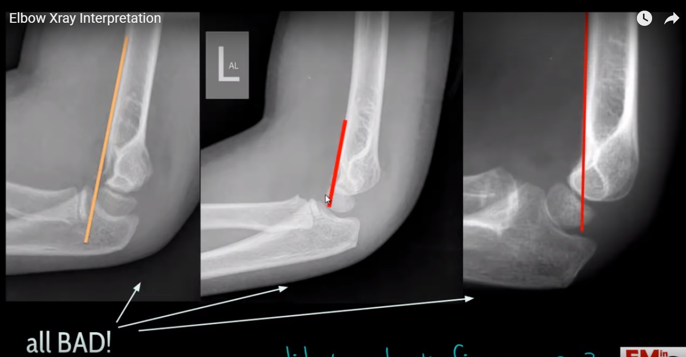
EMRad Radiologic Approach to the Pediatric Traumatic Elbow Xray Elbow X-Ray Normal — standard radiographic examination of the elbow includes anteroposterior (ap) and lateral views. Soft tissue areas, cortical margins, trabecular patterns, bony alignment, joint. — the elbow series is a set of radiographs taken to investigate elbow joint pathology, often in the context of trauma. On the lateral image there is often a visible triangle of low density lying. Elbow X-Ray Normal.
